IMAGE

Fig. 5.

ID
ZDB-IMAGE-250828-159
Source
Figures for Yu et al., 2025
Image
Figure Caption

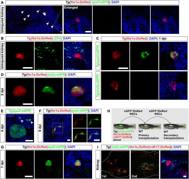
Fig. 5. Renewal of RSCs in adult kidneys.

(A) Confocal images of uninjured adult Tg(lhx1a:DsRed;eya2:eGFP) kidney sections showing lhx1a:DsRed+ RSC aggregates, with the cell aggregate (indicated by arrowheads) magnified for detailed observation. At this stage, RSC aggregates do not express eya2:eGFP. (B) EdU assay of uninjured Tg(lhx1a:DsRed) kidney sections. EdU was administered intraperitoneally every third day until kidney collection for analysis. Results show that most RSC aggregates are not labeled by EdU. (C) Confocal images of Tg(lhx1a:DsRed;eya2:eGFP) and Tg(lhx1a:DsRed;six2a:eGFP) kidney sections at 1 dpi, showing that the aggregates begin to express eya2:eGFP but not six2a:eGFP. Each RSC aggregate contains approximately 20 cells at this stage. (D) Confocal images of Tg(lhx1a:DsRed;six2a:eGFP) kidney sections at 2 dpi, showing the appearance of six2a:eGFP+ cells. By this stage, the RSC aggregates expand to approximately 50 cells. (E) Confocal images of Tg(eya2:eGFP) kidney sections at 4 dpi, indicating that nascent RSCs emerge. Arrowheads indicate individual RSCs. (F and G) Confocal images of Tg(lhx1a:DsRed;eya2:eGFP) kidney sections at 6 dpi (F) and 7 dpi (G), showing that nascent RSCs begin to express lhx1a and aggregate to form new cell aggregates. (H) Schematic of the serial transplantation assay. (I) Confocal images showing donor-derived nephrons (cdh17:DsRed+) and RSCs (six2a:eGFP+) in primary and secondary engrafted recipients. White dashed lines indicate the boundary of the kidney. Scale bars, 50 μm [(A) to (E) and (G) to (I)] and 10 μm (F).

Acknowledgments
This image is the copyrighted work of the attributed author or publisher, and ZFIN has permission only to display this image to its users. Additional permissions should be obtained from the applicable author or publisher of the image. Full text @ Sci Adv