IMAGE

Fig. 2

ID
ZDB-IMAGE-250814-27
Source
Figures for Han et al., 2025
Image
Figure Caption

Fig. 2

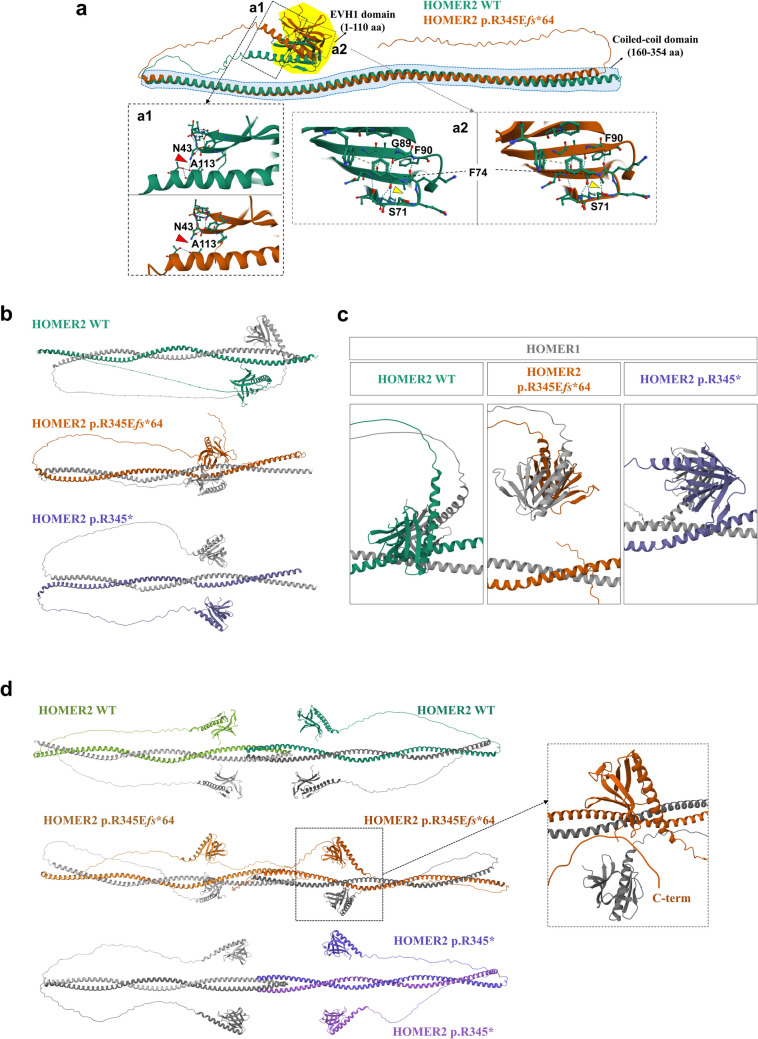
Predicted 3D structures of HOMER2 WT and HOMER2 p.R345Efs*64 using AlphaFold2. a Structural alignment of HOMER2 WT and HOMER2 p.R345Efs*64. The EVH1 domain is highlighted in the yellow area, and the coiled-coil (CC) domain is marked by the blue dashed line. a1 The hydrogen bond between N43 and A113 is disrupted in HOMER2 p.R345Efs*64 compared to HOMER2 WT. a2 The β-sheet structure in HOMER2 p.R345Efs*64 extends to F90, and a hydrogen bond forms between F74 and S71, which is absent in HOMER2 WT. b The structure of the HOMER1 [NP_004263.1] and HOMER2 dimer is shown. HOMER1 is depicted in gray, while HOMER2 WT, HOMER2 p.R345Efs*64, and HOMER2 p.R345* are represented in green, brown, and purple, respectively. The coiled-coil region of HOMER2, which can interact with other proteins, is located at amino acids 307–329 in the WT and is shifted to amino acids 275–297 in the HOMER2 variants. c Structural changes in the EVH1 domain were detected in the HOMER1 WT and HOMER2 variants dimer. The distance between each EVH1 domain of HOMER1 and HOMER2 p.R345Efs*64 was reduced (b) compared to the WT, which is shifted vertically. In HOMER1 WT and HOMER2 p.R345* dimer, the distance between each EVH1 was increased (b) and slightly shifted vertically. d The predicted tetramer structures were obtained using HOMER1 and HOMER2. Two HOMER1 proteins were combined with two HOMER2 WT, two HOMER2 p.R345Efs*64, or two HOMER2 p.R345* molecules. In the HOMER1 and HOMER2 p.R345Efs*64 tetramer, the location of the EVH1 domain of HOMER1 was altered, and the C-terminal of HOMER2 approached the EVH1 domain of HOMER2 (dashed black box). In the HOMER1 and HOMER2 p.R345* tetramer, HOMER1 and HOMER2 formed homodimers, and each homodimer formed a tetramer, unlike the other configurations. WT, wild-type; R, arginine; E, glutamate; fs, frameshift; EVH1, Ena/Vasp homology domain 1; F, phenylalanine; S, serine; N, asparagine; A, alanine

Acknowledgments
This image is the copyrighted work of the attributed author or publisher, and ZFIN has permission only to display this image to its users. Additional permissions should be obtained from the applicable author or publisher of the image. Full text @ J. Mol. Med.