IMAGE

Figure 5.

ID
ZDB-IMAGE-250705-23
Source
Figures for Abgueguen et al., 2025
Image
Figure Caption

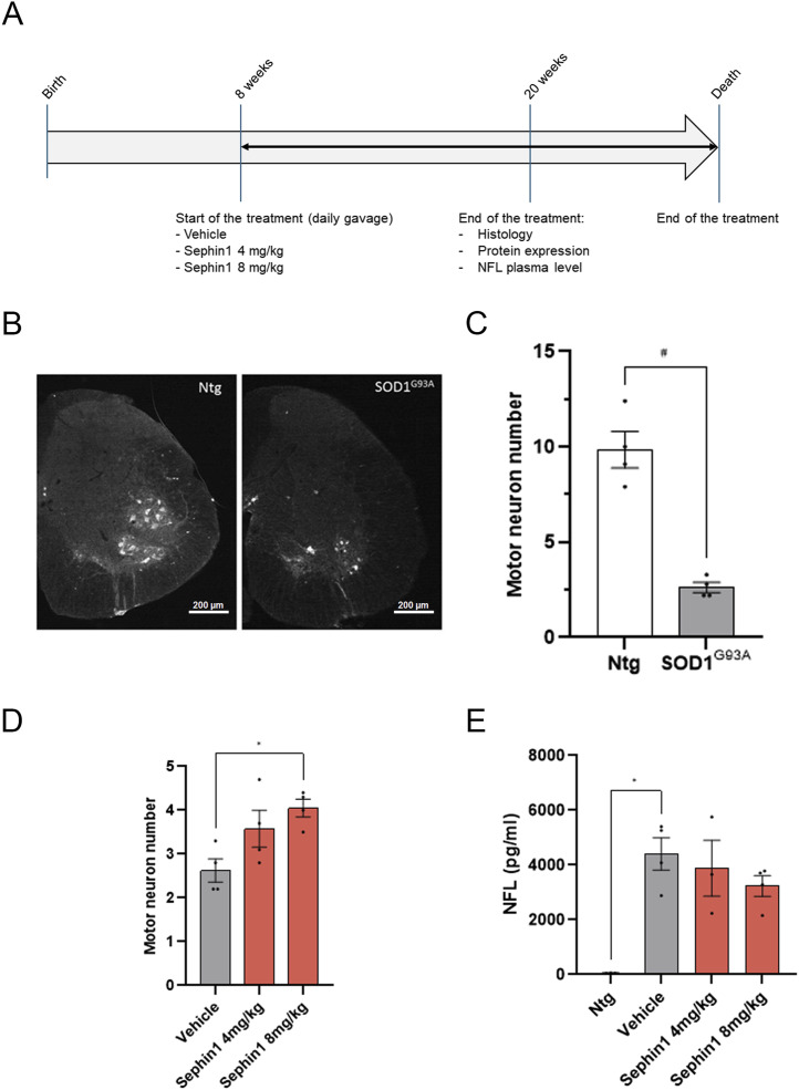
Figure 5. Sephin1 improves survival of spinal cord motor neurons from SOD1G93A female mice at 20 wk of age.

(A) Schematic of the experiment on female SOD1G93A mice. (B) Representative pictures of motor neurons labeled with ChAT antibody in hemisection of spinal cord of WT (Ntg) and SOD1G93A mice at 20 wk of age (magnification 4×). (C) Motor neuron number in spinal cord of 20 wk-old WT or SOD1G93A mice. (D) Motor neuron number in spinal cord of 20-wk-old SOD1G93A mice treated with vehicle, Sephin1 at 4 or 8 mg/kg per day. (E) Plasmatic NFL level in 20-wk-old WT or SOD1G93A mice treated with vehicle, Sephin1 at 4 or 8 mg/kg per day. #P < 0.05 Mann-Whitney test; *P < 0.05 versus vehicle Kruskal-Wallis test followed by Dunn’s multiple comparison test; mean ± SEM n = 3–5 mice per condition.

Acknowledgments
This image is the copyrighted work of the attributed author or publisher, and ZFIN has permission only to display this image to its users. Additional permissions should be obtained from the applicable author or publisher of the image. Full text @ Life Sci Alliance