IMAGE

Figure S1.

ID
ZDB-IMAGE-250705-18
Source
Figures for Abgueguen et al., 2025
Image
Figure Caption

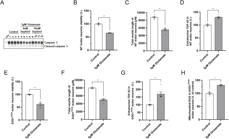
Figure S1. Glutamate intoxication decreases cell viability, neurite network integrity, and increases extranuclear TDP-43 in primary motor neurons from WT and SOD1G93A rats.

(A) Representative picture of immunoblot obtained by WES automated WB apparatus for caspase 3 and cleaved caspase 3 proteins in primary WT rat motor neurons intoxicated with glutamate and treated with DMSO or Sephin1 50 nM. (B) Cell viability in control and glutamate intoxicated primary WT rat motor neurons. (C) Total neurite length in control and glutamate intoxicated primary WT rat motor neurons. (D) Percentage of extranuclear TDP-43 in control and glutamate intoxicated primary WT rat motor neurons. (E) Cell viability in control and glutamate intoxicated primary SOD1G93A rat motor neurons. (F) Total neurite length in control and glutamate intoxicated primary SOD1G93A rat motor neurons. (G) Percentage of extranuclear TDP-43 in control and glutamate intoxicated primary SOD1G93A rat motor neurons. (H) Mitochondrial ROS level in control and glutamate intoxicated primary SOD1G93A rat motor neurons. *P < 0.05, **P < 0.01 Mann-Whitney test, mean ± SEM n = 4–6 replicates per condition.

Acknowledgments
This image is the copyrighted work of the attributed author or publisher, and ZFIN has permission only to display this image to its users. Additional permissions should be obtained from the applicable author or publisher of the image. Full text @ Life Sci Alliance