IMAGE

Figure 1

ID
ZDB-IMAGE-250701-38
Source
Figures for Rosa et al., 2025
Image
Figure Caption

Figure 1

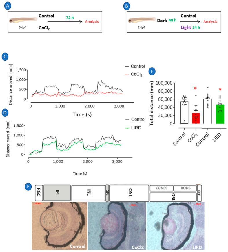
Retinopathy in zebrafish larvae. Retinopathy was induced in 3 dpf larvae through exposure to E2 0.5x medium at 28 °C with 0.5 mM cobalt chloride (CoCl2) for 72 h (A). Independent group of 3 dpf larvae was exposed to intense light at 8000 lux for 24 h (LIRD) (B). Larvae maintained at 28 °C in E2 0.5x medium only and under natural light were considered negative controls. Immediately after stimulation, all groups of larvae were evaluated for their visual–motor response (C,D) and distance traveled (E) and were subsequently killed and processed for histological analysis and H&E staining (F). * p < 0.05 compared to control group. Bars represent mean ± SEM. Dots represent individual data points. RGL: retinal ganglion layer; IPL: inner plexiform layer; INL: inner nuclear layer; OPL: outer plexiform layer; ONL: outer plexiform layer; OSL: outer segment layer; RPE: retinal pigment epithelium.

Acknowledgments
This image is the copyrighted work of the attributed author or publisher, and ZFIN has permission only to display this image to its users. Additional permissions should be obtained from the applicable author or publisher of the image. Full text @ Pharmaceuticals (Basel)