IMAGE

Fig. 2

ID
ZDB-IMAGE-250526-61
Source
Figures for Shi et al., 2025
Image
Figure Caption

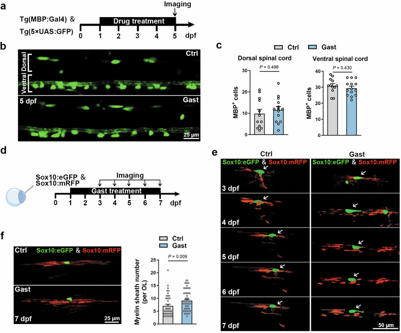
Fig. 2 Gastrodin promotes myelin sheath formation.

a Schematic of Tg(MBP:Gal4) & Tg(5×UAS:GFP) imaging pipeline. b Representative images showing lateral views of the spinal cords at 5 dpf. c Quantification of GFP+ cells number in the ventral or dorsal spinal cord (n = 12–14 zebrafish). d Schematic of DNA microinjection and imaging pipeline. e Time-lapse imaging of myelin sheath development of a single OL from 3 dpf to 7 dpf. f Representative images showing myelin sheaths formed by a single OL at 7 dpf and quantification of myelin sheath number per OL (n = 42–60 OLs). Data are represented as mean ± SEM, unpaired Student’s t test in c and f.

Acknowledgments
This image is the copyrighted work of the attributed author or publisher, and ZFIN has permission only to display this image to its users. Additional permissions should be obtained from the applicable author or publisher of the image. Full text @ Acta Pharmacol. Sin.