
|
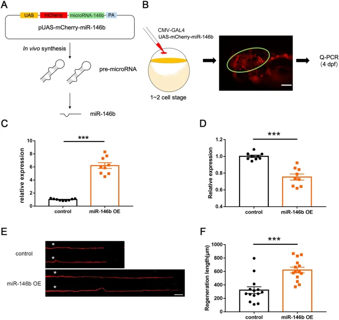
Fig. 4 Reprogramming miR-146b-snphb signaling promotes M-cell axon regeneration. A Schematic of the in vivo overexpression system of miR-146b. B Validation of vector-based miR-146b overexpression. The circle indicates neurons in the brain that express fluorescent signals. C MiR-146b levels were significantly increased by vector-based expression in vivo. D Quantitative RT-PCR showed that the expression of snphb was significantly reduced at 10 hpf in embryos overexpressing miR-146b. E, F Representative images of control (UAS-mCherry) and miR-146b OE axon regeneration results. Statistical results showed that miR-146b OE enhanced axon regeneration in zebrafish M-cells. Control: 322.1 ± 49.65 μm; miR-146b OE: 622.2 ± 42.48 μm. White asterisk denotes the injury site. Scale bar, 50 μm, *** P < 0.001, error bars indicate SEM.