
Image
|
Figure Caption
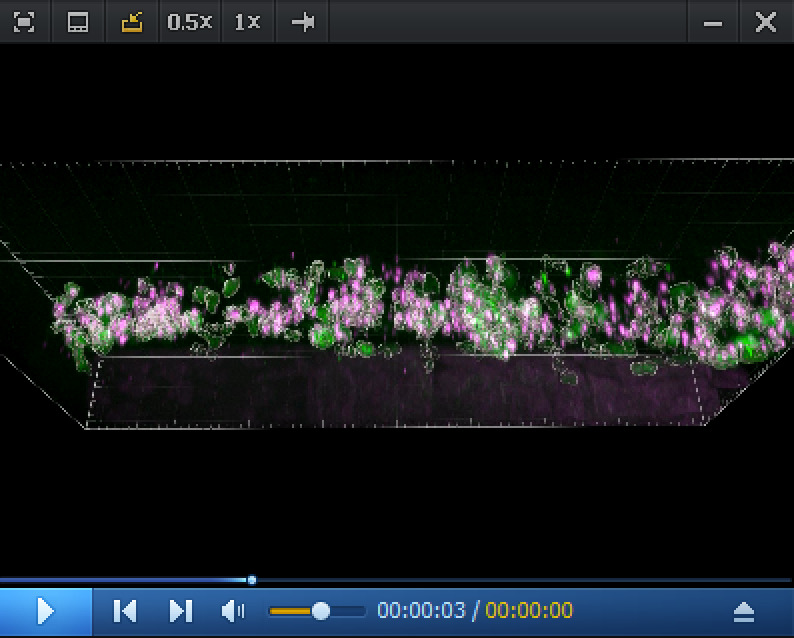
Video 3. Imaris 3D rendering and colocalization of RNAscope spots and hematopoietic stem and progenitor cells (HSPCs) in the CHT region.
3D reconstitution generated from a spinning disk confocal z-stack obtained from a 5 dpf
Acknowledgments
This image is the copyrighted work of the attributed author or publisher, and
ZFIN has permission only to display this image to its users.
Additional permissions should be obtained from the applicable author or publisher of the image.
Full text @ Bio Protoc