
Image
|
Figure Caption
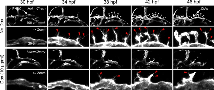
Fig. 5 Activated Wnt/β-catenin signaling in NPCs decreases tip cell migration. Lateral still frames from time-lapse confocal microscopy of gfap:rtTA, TRE:β-cat*-EGFP larvae treated with indicated Dox concentrations. Snapshots are at 4-h intervals over 16 h of acquisition beginning at the onset of CNS angiogenesis (30 hpf). Magnified frames (4 × Zoom) highlight the hindbrain region bounded ventrally by the primordial hindbrain channels. Stereotypical tip cells are indicated by white asterisks. Stereotypical filipodia are indicated by red arrowheads. See supplementary videos 4, 5 for more detail.
Acknowledgments
This image is the copyrighted work of the attributed author or publisher, and
ZFIN has permission only to display this image to its users.
Additional permissions should be obtained from the applicable author or publisher of the image.
Full text @ Sci. Rep.