IMAGE

Fig. 1

ID
ZDB-IMAGE-250225-73
Source
Figures for Goh et al., 2025
Image
Figure Caption

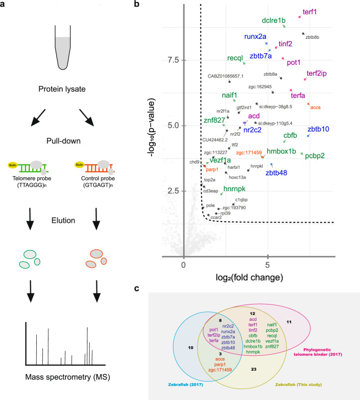
Fig. 1 Telomere pull-down with the zebrafish cell line.

a Schematic diagram showing an overview of the telomere pull-down experiment using nuclear lysate from the zebrafish fin fibroblast BRF41 cell line. b Volcano plot visualizing the results obtained by label-free quantification of the bound proteins. The pull-down experiment was conducted in quadruplicate using either concatenated telomeric TTAGGG or a control scrambled GTGAGT oligonucleotide as bait. The results were log-transformed and plotted on the x-axis as log2(fold change) and on the y-axis as -log10(p-value). The protein enrichment threshold was set at a fold change >2 and a p-value < 0.05 (Welch’s t test) with c = 0.05. Enriched proteins were annotated with their gene names and color-coded: shelterin subunits (magenta), proteins that were previously found in the study by Kappei et al. to be enriched in zebrafish (orange), and telomere-binding proteins previously identified as phylogenetically conserved in other species (green), which were also enriched in zebrafish (blue). c Comparison of the previous telomere zebrafish pull-down, the phylogenetic conserved binders and the candidates from the current study.

Acknowledgments
This image is the copyrighted work of the attributed author or publisher, and ZFIN has permission only to display this image to its users. Additional permissions should be obtained from the applicable author or publisher of the image. Full text @ Commun Biol