IMAGE

Fig. 7

ID
ZDB-IMAGE-250219-61
Source
Figures for Dharmadhikari et al., 2025
Image
Figure Caption

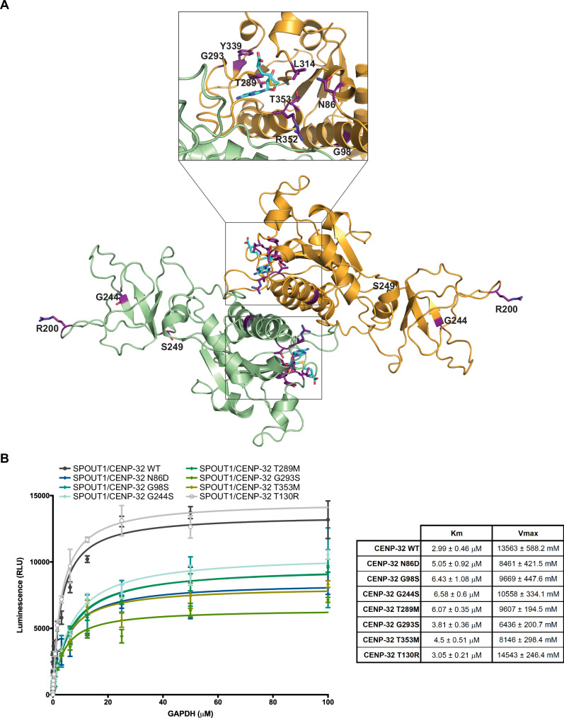
Fig. 7 SPOUT1/CENP-32 patient variants show decreased methyltransferase activity.

A Mapping of the SPOUT1/CENP-32 variants in the SPOUT1/CENP-32 71-376 SAH-bound structure. Variant residues are highlighted in purple. B Methyltransferase assay to determine the Km and Vmax of SPOUT1/CENP-32 WT and the variants in the presence of constant SAM (20 μM) and increasing amounts of a GAPDH mRNA hairpin as substrate (GCCCCCUCUGCUGAUGCCCCCAUGUUCGUCAUGGGUGUGAA; 0 – 100 μM; left panel). Km and Vmax values for all SPOUT1/CENP-32 proteins are depicted in the table (right panel). R square values are as follows: SPOUT1/CENP-32 WT – 0.79 (n = 10), SPOUT1/CENP-32 N86D – 0.83 (n = 6), SPOUT1/CENP32 G98S – 0.87 (n = 6), SPOUT1/CENP-32 G244S – 0.93 (n = 10), SPOUT1/CENP-32 T289M – 0.97 (n = 10), SPOUT1/CENP-32 G293S – 0.93 (n = 10), SPOUT1/CENP-32 T353M – 0.9 (n = 10), SPOUT1/CENP-32 T130R – 0.98 (n = 4). Data from three independent biological replicates, mean ± SD. Source data are provided as a Source Data file.

Acknowledgments
This image is the copyrighted work of the attributed author or publisher, and ZFIN has permission only to display this image to its users. Additional permissions should be obtained from the applicable author or publisher of the image. Full text @ Nat. Commun.