
|
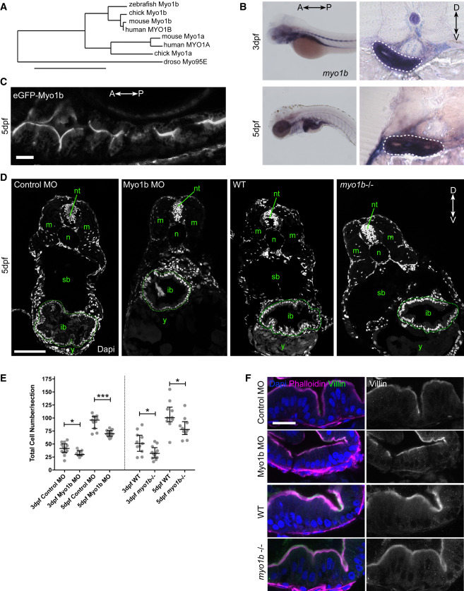
Fig. 4 Myo1b localizes apically in the zebrafish gut epithelium but is not involved in epithelial cell differentiation (A) Phylogenetic tree based on protein sequence of zebrafish, chick, human, and mouse Myo1b and Myo1a and Drosophila Myo95E. (B) In situ hybridization for myo1b transcripts on 3- and 5-dpf zebrafish larvae whole mounts (left panels) and cross-sections at the level of the intestinal bulb (right panels). On sections, the forming intestinal bulb is circled with white dashed lines. (C) Live, longitudinal (anteroposterior axis) confocal section of the intestinal bulb of a 5-dpf zebrafish larva expressing the transcription activator KalT4 driving the expression of the eGFP-Myo1b transgene under the control of an upstream activating sequence in the gut epithelium. Scale bar, 30 μm. (D) Confocal imaging of zebrafish section sections stained with DAPI of 5-dpf larvae injected with control and Myo1b MO, and 5-dpf WT and myo1b−/− larvae. ib, intestinal bulb (circled with dashed lines); m, muscles; n, notochord; nt, neural tube; sb, swim bladder; y, yolk. Scale bar, 100 μm. (E) Quantifications from DAPI-stained sections of the total number of cells per section at 3 and 5 dpf in the four conditions. Data are presented as median and interquartile range; Mann-Whitney test, ∗p < 0.05, ∗∗∗p < 0.001. (F) Imaging of zebrafish larval sections of the intestinal bulb at 5 dpf in the four conditions stained for the microvilli marker Villin, F-actin (phalloidin), and DAPI. Scale bar, 20 μm.