
Fig. 3
- ID
- ZDB-IMAGE-250110-32
- Genes
- Publication
- Guggeri et al., 2024 - Follistatin like-1 (Fstl1) regulates adipose tissue development in zebrafish
- All Figures
- Figures for Guggeri et al., 2024
|
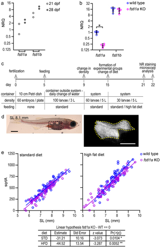
Fig. 3 fstl1a KO larvae have less adipose tissue than wild-type larvae. a) Dot plot showing the qPCR results to assess the levels of expression of fstl1a and fstl1b in the isolated abdominal fat deposit of 21 and 28 dpf wild-type larvae (the mean fstl1a level at 21dpf was used as reference; samples from two larvae). b) Dot plot showing the qPCR results for fstl1a and fstl1b in the abdominal region of 21 dpf wild-type and fstl1a KO larvae. Five larvae were analyzed in each condition. Plot: individual NRQ values and mean ± CI (95%). (*) p = 0.012, Mann–Whitney test. c) Schematic representation of the protocol followed to perform the diet experiments. d) Examples of the panoramic bright field and epifluorescence images of Nile Red stained larvae used to quantify the standard length (SL) and extension (area) of adipose tissue. Scale bar: 1 mm (left image); 0.5 mm (right image). e) Dot plots comparing the square root of the area of the adipose tissue vs SL of wild-type and fstl1a KO larvae in standard and high-fat diets. The straight lines are the least squares fit to the pooled data from three experiments. The statistical comparison was made using a linear mixed model fit and Tuckey contrasts (bottom table) (see the complete analysis in Figure S3).