
Image
|
Figure Caption
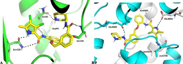
Fig. 3 Predicted binding mode of (R)-3 (in yellow sticks) in the (a) ATP-binding site of the human topoisomerase IIα (PDB entry: 4R1F, in green cartoon), and (b) allosteric binding site at the Hsp90β C-terminal domain (PDB entry: 5FWK, protomer A in grey cartoon, protomer B in cyan cartoon). Hydrogen bonds between the protein and (R)-3 are presented as black dashed lines.
Acknowledgments
This image is the copyrighted work of the attributed author or publisher, and
ZFIN has permission only to display this image to its users.
Additional permissions should be obtained from the applicable author or publisher of the image.
Full text @ Bioorg. Chem.