
Image
|
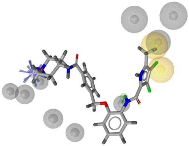
Figure Caption
Fig. 2 Overlay of compound 3 with the structure-based pharmacophore model derived from the most representative molecular dynamics binding mode of inhibitor in the C-terminal domain binding site of the Hsp90β dimer [28] (PDB entry: 5FWK). Blue star represents a positively ionizable pharmacophore feature, green arrow a hydrogen bond donor pharmacophore feature, yellow spheres hydrophobic pharmacophore feature and grey spheres represent excluded volumes.
Acknowledgments
This image is the copyrighted work of the attributed author or publisher, and
ZFIN has permission only to display this image to its users.
Additional permissions should be obtained from the applicable author or publisher of the image.
Full text @ Bioorg. Chem.