IMAGE

Fig. 2.

ID
ZDB-IMAGE-241218-47
Source
Figures for Desingu Rajan et al., 2024
Image
Figure Caption

Fig. 2.

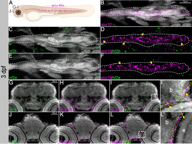
nf2a/b is expressed in Schwann cells and meninges. (A) Schematic representing the posterior lateral line Schwann cells (pLLn SCs; white dashed line box) and sectioned regions (black dashed lines) at 3 dpf. (B-F) Multiplexed fluorescent mRNA in situ hybridizations by HCR on whole-mount embryos reveal expression of nf2b, nf2a and sox10 in a Schwann cell cluster at 3 dpf. Yellow arrows indicate cells that have both sox10 and nf2 expression. White dashed lines indicate pLLn SCs expressing sox10. (G-I,J-L) Multiplexed fluorescent mRNA in situ hybridizations by HCR on cryo-sectioned embryos reveal expression of nf2b, nf2a and foxc1b in the cranial meninges at 3 dpf. (I′,L′) Magnified images of the regions in the white dashed line boxes in I and L, respectively. Yellow dashed lines mark the meninges layer in close proximity to the brain; yellow arrowheads indicate cells that have both foxc1b and nf2 expression. Images are representative of n=7 embryos. Images in B-F represent confocal planes of 5 μm thickness. G-L are histological sections of 12 μm thickness imaged with Apotome. DAPI (gray) was used as a counterstain. b, brain; e, eye.

Acknowledgments
This image is the copyrighted work of the attributed author or publisher, and ZFIN has permission only to display this image to its users. Additional permissions should be obtained from the applicable author or publisher of the image. Full text @ Dis. Model. Mech.