
IMAGE
Figure S5.
- ID
- ZDB-IMAGE-241021-32
- Publication
- El-Daher et al., 2024 - Microglia are essential for tissue contraction in wound closure after brain injury in zebrafish larvae
- All Figures
- Figures for El-Daher et al., 2024
Image
|
Figure Caption
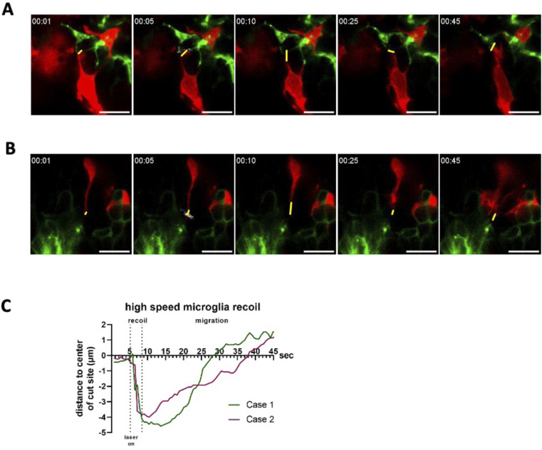
Figure S5. Single-plane high temporal resolution imaging of recoiling microglial contacts after laser cutting.
Acknowledgments
This image is the copyrighted work of the attributed author or publisher, and
ZFIN has permission only to display this image to its users.
Additional permissions should be obtained from the applicable author or publisher of the image.
Full text @ Life Sci Alliance