
|
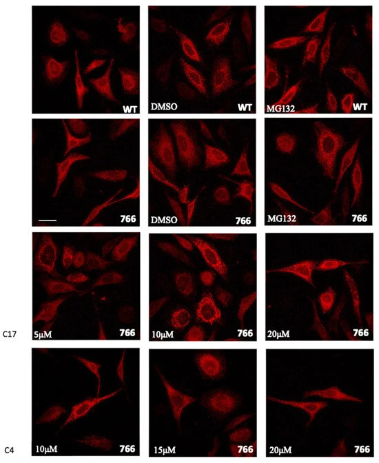
Fig. 6 Cellular expression and localization of WT and mutant SERCA1 protein in HeLa cells. Cells were transfected with WT or with S766F mutated SERCA1 cDNAs, as indicated. Sixteen hours after transfection, MG132 (10 μM final concentration dissolved in DMSO), DMSO (its vehicle 0,1%) and different concentrations of C17 (5 μM, 10 μM, 20 μM) or C4 (10 μM, 15 μM, 20 μM) CFTR correctors, were added and cells were incubated for 8h. Transfected and treated cells were immunolabelled with monoclonal antibodies to SERCA1 and then incubated with the Alexa 568 red fluorescence secondary antibody. Images were recorded at the same setting conditions and magnification (scale bar 50 µm).