
Fig. 1
- ID
- ZDB-IMAGE-240912-52
- Genes
- Antibodies
- Publication
- Huang et al., 2024 - mTORC1 mediates the expansion of hematopoietic stem and progenitor cells through ribosome biogenesis protein Urb2 in zebrafish
- All Figures
- Figures for Huang et al., 2024
|
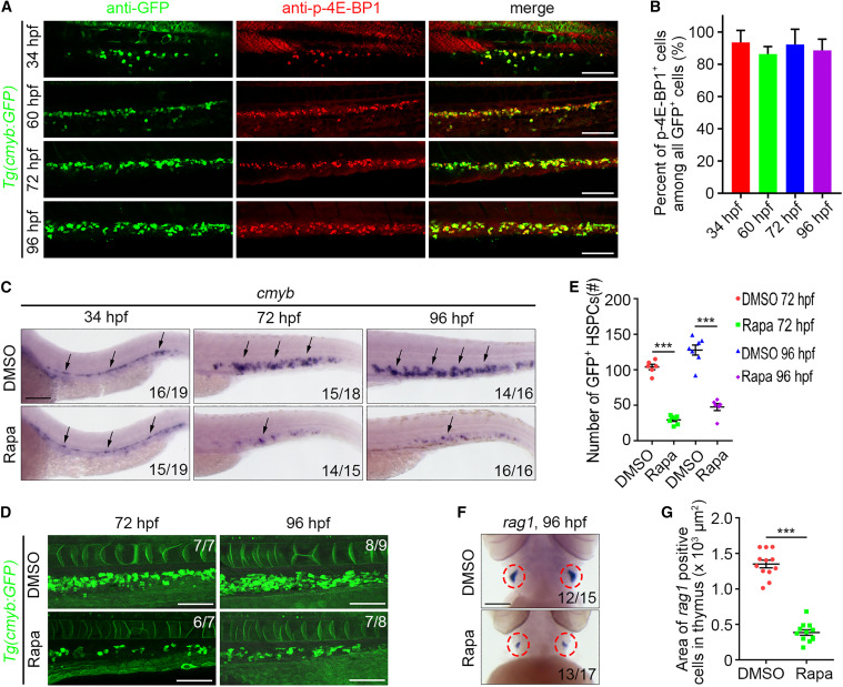
Fig. 1 mTORC1 signaling is required for HSPC development in zebrafish (A) Single-optical section images showing GFP and p-4E-BP1 antibody staining under Tg(cmyb:GFP) background at 34 , 60 , 72 , and 96 hpf. (B) Quantification of the percent of p-4E-BP1+ cells among all GFP+ cells. (C) WISH images showing the cmyb expression at 34 , 72 , and 96 hpf. Arrows indicate AGM at 34 hpf and CHT at 72 and 96 hpf. (D) Confocal projection images showing the cmyb:GFP expression in CHT at 72 and 96 hpf. (E) Quantification of the number of GFP+ HSPCs. ∗∗∗p < 0.001 on unpaired two-tailed t test (n = number of total embryos from three independent experiments). (F) WISH images showing the rag1 expression at 96 hpf. (G) Quantification of the area of rag1+ cells in the thymus. Circles indicate the thymus. ∗∗∗p < 0.001 on unpaired two-tailed t test (n = number of total embryos from three independent experiments). Error bars represent SEM. Scale bars: 100 μm. Rapa, rapamycin.