
Fig. 5
- ID
- ZDB-IMAGE-240903-67
- Genes
- Publication
- Harboe et al., 2024 - The metalloproteinase PAPP-A is required for IGF-dependent chondrocyte differentiation and organization
- All Figures
- Figures for Harboe et al., 2024
|
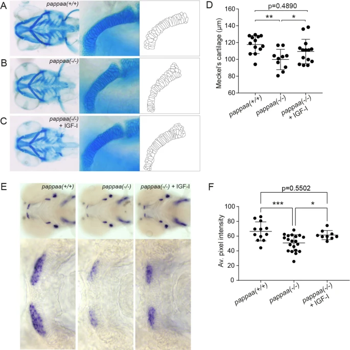
Fig. 5 Exogenous IGF-I restores normal morphology of Meckel’s cartilage and expression of col10a1a. (A–C) 3 dpf pappaa knockout larvae were transferred to E3 buffer with or without 30 ng/mL IGF-I. At 4 dpf, the larvae were terminated, Alcian blue-stained and the length of Meckel’s cartilage was determined. Representative ventral images of nontreated and IGF-I-treated larvae, corresponding magnifications of Meckel’s cartilage, and outlines of the chondrocytes are shown. (D) Length of Meckel’s cartilage for non-treated wild-type (n = 13) or knockout (n = 10) pappaa, and IGF-I-treated knockout pappaa (n = 14). (E) Pappaa knockout larvae were treated as described in (A), and stained for col10a1a by whole mount in situ hybridization. Representative ventral images and magnifications of Meckel’s cartilage for are shown. (F) Average intensity of the col10a1a staining in Meckel’s cartilage. Data are from three independent experiments, shown as mean ± SD and analyzed by one-way ANOVA. * p < 0.05; ** p < 0.01; *** p < 0.001; **** p < 0.0001