IMAGE

Fig. 4

ID
ZDB-IMAGE-240902-42
Source
Figures for Arroyo et al., 2024
Image
Figure Caption

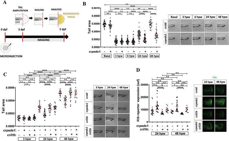
Fig. 4 Peds1 deficiency hampers tissue regeneration.

A Schematic of the experimental procedure used for tail injury and regeneration assays. Three dpf larvae previously microinjected with std or peds1 crRNA/Cas9 complexes alone or in combination with cril1b were tail fin amputated and regeneration was evaluated by measuring regenerated fin areas up to 5 dpf (yellow). B, C Measurement of regenerated fin areas and representative images (brightfield channel). D Analysis and representative images of Il1b fluorescent reporter expression levels (green channel; Tg(il1b:GFP-F)ump3) by microscopy fluorescence. Each point represents one larva and the mean ± SEM of each group is also shown. P-values were calculated using one-way ANOVA and Tukey’s multiple range. n.s, not significant, *p ≤ 0.05, **p ≤ 0.01, ***p ≤ 0.001, ****p ≤ 0.001. auf arbitrary units of fluorescence.

Acknowledgments
This image is the copyrighted work of the attributed author or publisher, and ZFIN has permission only to display this image to its users. Additional permissions should be obtained from the applicable author or publisher of the image. Full text @ Cell Death Discov