IMAGE

Figure 3

ID
ZDB-IMAGE-240729-253
Source
Figures for Filippini et al., 2024
Image
Figure Caption

Figure 3

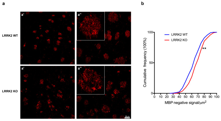
LRRK2 KO mouse brains displayed alterations of MBP+ striosomes. (a) Maximum intensity Z-projection confocal images of 13-month-old LRRK2 WT and KO mice immunostained for MBP. LRRK2 WT: (a’) Z = 3 mm, (a’’) Z = 4 mm; LRRK2 KO: (a’) Z = 3 mm, (a’’) Z = 2.33 mm. Scale bars 20 µm. (b) Cumulative frequency distributions of MBP negative signal in LRRK2 WT and LRRK2 KO mice (LRRK2 WT = 249 striosomes and LRRK2 KO = 242 striosomes; ** p < 0.01, Kolmogorov–Smirnov test, n = 3 animals per group).

Acknowledgments
This image is the copyrighted work of the attributed author or publisher, and ZFIN has permission only to display this image to its users. Additional permissions should be obtained from the applicable author or publisher of the image. Full text @ Biomolecules