
Fig. 3
- ID
- ZDB-IMAGE-240620-75
- Publication
- Du et al., 2024 - Lapatinib combined with doxorubicin causes dose-dependent cardiotoxicity partially through activating the p38MAPK signaling pathway in zebrafish embryos
- All Figures
- Figures for Du et al., 2024
|
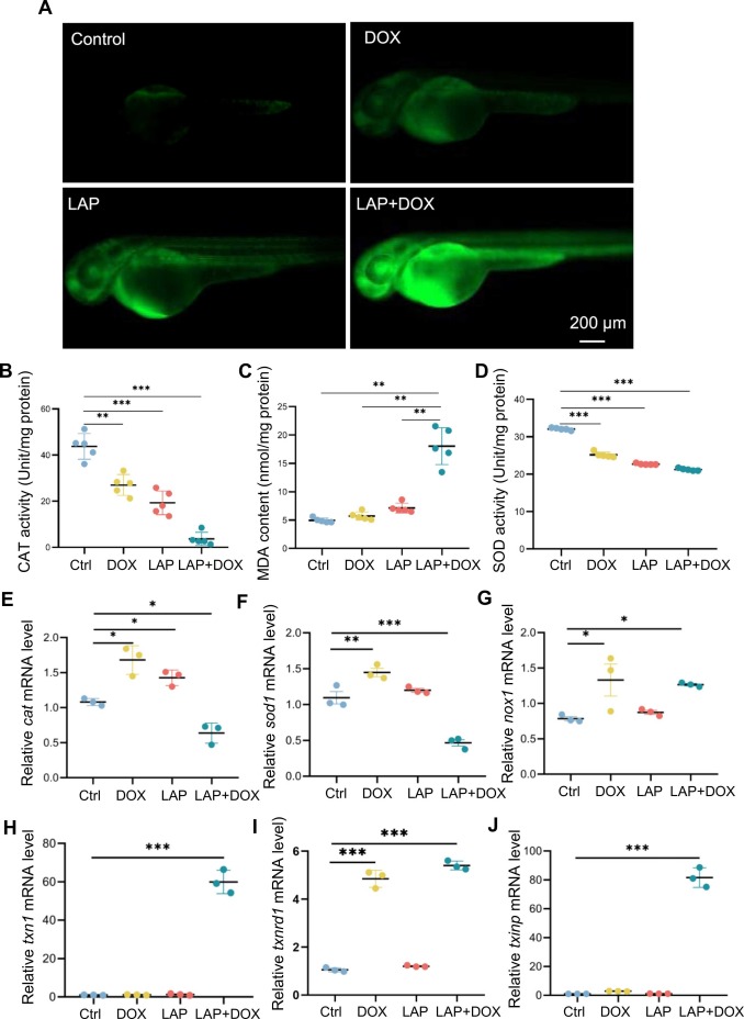
Fig. 3 Oxidative stress was increased in the combined LAP and DOX exposure-induced cardiotoxicity model. (A) Representative fluorescence images of ROS staining in the combined LAP and DOX exposure-induced cardiotoxicity model compared to embryos exposed to LAP or DOX alone or DMSO-treated control embryos. (B-D) Quantification of other ROS-related indices, including catalase (CAT) (B), superoxide dismutase (SOD) (C) and malondialdehyde (MDA) (D), in the LAP and DOX combination-induced cardiotoxicity model compared to embryos exposed to LAP or DOX alone or DMSO-treated control embryos. N=5, one-way ANOVA; (E-J) Quantitative RT-PCR analysis of ROS-related gene expression in the LAP and DOX combination-induced cardiotoxicity model compared to embryos exposed to LAP or DOX alone or DMSO-treated control embryos. N=3 biological repeats. one-way ANOVA. *P<0.05, **P<0.01, ***P<0.001.