IMAGE

Fig. 4

ID
ZDB-IMAGE-240612-23
Source
Figures for Akam-Baxter et al., 2024
Image
Figure Caption

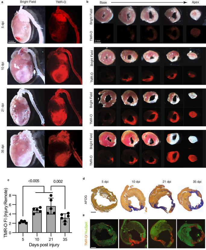
Fig. 4 Collagen oxidation and cross-linking in the infarcted mouse heart.

a The hearts of adult mice injected with TMR-O (2 nmol/g, IV) at 5–35 days after myocardial infarction (MI) are shown in their long axis. TMR-O fluorescence in the infarct is, qualitatively, already high at day 10, (n = 5 except for 35 dpi where n = 6). b Sectioning of the heart into 1 mm thick short axis slices confirms that the TMR-O signal is arising from the infarcted myocardium and is patchy but substantial by day 10 (n = 5 except for 35 dpi where n = 6). c Time course of TMR-O fluorescence in murine infarcts(n = 5 except for 35 dpi where n = 6). The oxidation and cross linking of collagen is already significant by day 10, peaks at day 21 and subsequently decreases. Data are means ± SD of 5-6 measurements where each data point represents one mouse. Source data are provided in the Source Data file. P-values are shown where significant, one-way ANOVA with post hoc comparisons, two tailed. d, e AFOG-stained histology sections at the mid-ventricular level show that the collagen-rich injury area (blue) colocalizes well with the TMR-O signal on fluorescence microscopy (n = 5 except for 35 dpi where n = 6). Healthy myocardium is stained with a green, fluorescent nuclear stain (NucSpot). Scale bars = 1 mm.

Acknowledgments
This image is the copyrighted work of the attributed author or publisher, and ZFIN has permission only to display this image to its users. Additional permissions should be obtained from the applicable author or publisher of the image. Full text @ Nat. Commun.