
|
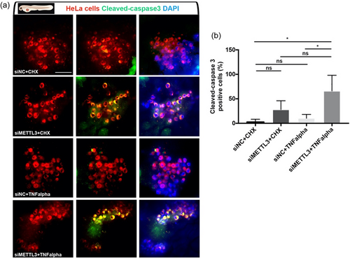
Fig. 7 Knockdown of METTL3 synergizes with TNF-α to promote apoptosis of HeLa cells in vivo (a) Representative confocal microscope images of anti-cleaved-caspase-3 (green) staining of 3 dpf (1 dpi) zebrafish larvae xenografted with HeLa cells (red) at 2 dpf, treated with negative control siRNA (si-NC) + CHX, si-METTL3 + CHX, si-NC + TNF-α or si-METTL3 + TNF-α. (b) Graph showing the percentage of cleaved-caspase3 positive cells in each treatment. Bars represent the average percentage of apoptotic cells counted in each z-stack slice divided by the number of DiL+, DAPI+ nuclei in (a). Larvae were counterstained for DAPI. Scale bars 50 μm.