IMAGE

Fig. 1.

ID
ZDB-IMAGE-240501-18
Source
Figures for Sharkova et al., 2024
Image
Figure Caption

Fig. 1.

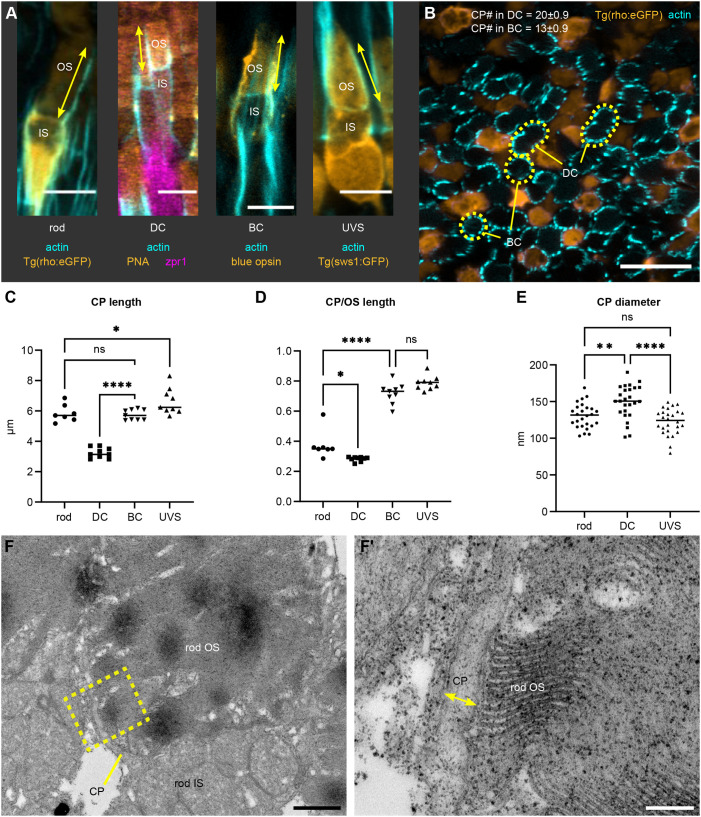
CP parameters for photoreceptor subtypes in the juvenile zebrafish retina. (A) Confocal images of 1 mpf retinas stained with phalloidin (cyan). Shown are a dark-adapted rod [DA; Tg(rho:eGFP)], light-adapted double cone (DC) [LA wild-type (WT) stained with PNA and zpr1], blue cone (BC) (LA WT stained with anti-blue opsin) and UV-sensitive cone (UVS) [LA Tg(sws1:GFP)]; CP length is indicated by arrows. (B) Sagittal section through a 1 mpf DA Tg(rho:eGFP) retina (rods in orange) labelled with phalloidin (cyan). BC and DC OSs are outlined. For CP number, mean±s.d. are shown; n=5 fish. (C,D) Graphs displaying CP length (C) and CP length relative to the OS length (D) for LA DC, LA BC, LA UVS cones, and DA rods; number of fish n=9 (DC, BC, UVS), n=7 (rod). (E) Graph showing CP diameter measured in rods, DC, and UVS cones in TEM images of 1 mpf WT retina, with individual measurements plotted; number of fish n=5. In C–E, the line highlights the median. ns, not significant (P>0.05), *P<0.05, **P<0.01, ****P<0.0001 (one-way ANOVA with Tukey's test). (F) Example of TEM imaging used for measuring CP diameter. Lower magnification image showing the rod OS, IS and a CP, with a yellow contour indicating the area in F′, where CP diameter is labelled. Scale bars: 5 µm (A), 10 µm (B), 1 µm (F), 200 nm (F′).

Acknowledgments
This image is the copyrighted work of the attributed author or publisher, and ZFIN has permission only to display this image to its users. Additional permissions should be obtained from the applicable author or publisher of the image. Full text @ J. Cell Sci.