IMAGE

Figure 4

ID
ZDB-IMAGE-240429-34
Source
Figures for Brenet et al., 2024
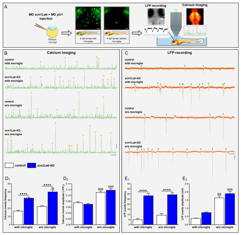
Image
Figure Caption

Figure 4

Microglia depletion increases neuron hyperactivity in scn1Lab-KD larvae. (A) Scheme of the experimental setup. (B) Calcium imaging of Ca++ uptake events recorded over a 20-min period in 4 dpf WT (N = 11), scn1Lab-KD (N = 16), microglia-depleted WT (N = 9) and microglia-depleted scn1Lab-KD larvae (N = 9). (C) LFP recording over a 20-min period of neuron activity in 4 dpf WT (N = 5), scn1Lab-KD (N = 5), microglia-depleted WT (N = 5) and microglia-depleted scn1Lab-KD larvae (N = 5). Scale bars of calcium traces: 0.05 ΔF/F0 and 1 min; and that of LFP traces: 0.5 mV and 1 min. Quantification of the frequency (D1) and amplitude (D2) of calcium events with intensity > 0.04 ΔF/F0, recorded during a 1-h period in 4 dpf not-injected (white) and scn1Lab-KD larvae (blue). (E). Quantification of the frequency (E1) and amplitude (E2) of LFP traces with downwards depolarization > 0.3 mV and lasting > 100 ms, recorded during a 1-h period in 4 dpf not injected (white) and scn1Lab-KD larvae (blue). N = number of larvae. All data are represented as the mean ± sem. ****, p < 0.0001: indicates a statistically significant difference between control and scn1Lab-KD larvae. $$, p < 0.01; $$$, p < 0.001; $$$$, p < 0.0001: indicates a statistically significant difference between larvae with or without microglia. p-values were determined using two-way ANOVA with Tukey post-tests.

Acknowledgments
This image is the copyrighted work of the attributed author or publisher, and ZFIN has permission only to display this image to its users. Additional permissions should be obtained from the applicable author or publisher of the image. Full text @ Cells