
|
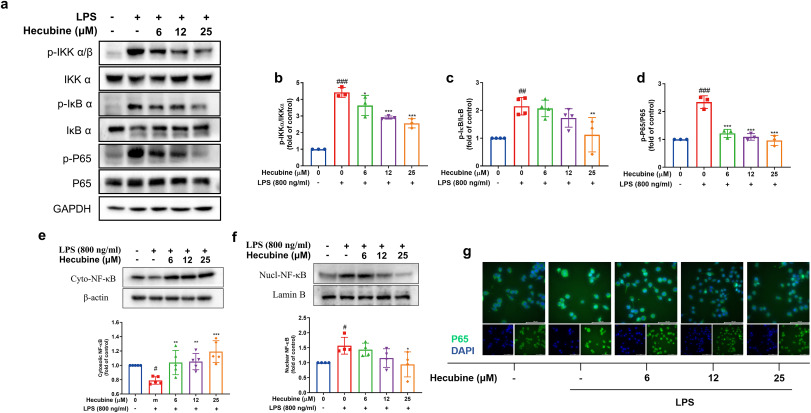
Fig. 6 Hecubine suppressed LPS-triggered activation of the NF-κB p65 pathway in BV-2 cells. Cells were pretreated with Hecubine for 1 h and then stimulated with/without LPS. (a–d) The levels of total/phosphorylated IKKα/β (b), IκB α (c), and P65 (d) were measured via Western blot (b, n = 3; c, n = 4; d, n = 3). (e–f) The expression levels of total NF-κB p65 in the cytoplasmic fraction (n = 5) (e) and nuclear fraction (n = 4) (f) were quantified by Western blot. (g) The nuclear translocation of p65 was evaluated by immunofluorescence analysis. Cells were stained with anti-p65 antibody (green) and DAPI (blue). – and + represent the absence and presence of LPS (800 ng/ml), respectively. The data are presented as the mean ± SD of three independent experiments. *p < 0.05, **p < 0.01, and ***p < 0.001 vs. LPS-treated group; ###p < 0.001 vs. control; One-way ANOVA with Dunnett’s multiple comparisons test. (For interpretation of the references to colour in this figure legend, the reader is referred to the Web version of this article.)