IMAGE

Fig. 3

ID
ZDB-IMAGE-240401-34
Source
Figures for Topi et al., 2024
Image
Figure Caption

Fig. 3

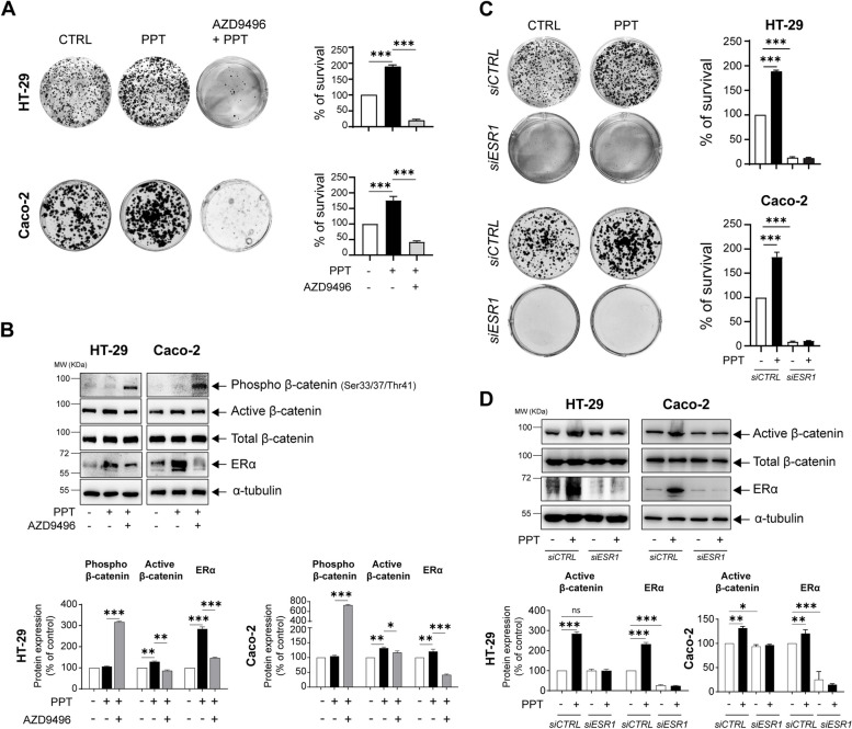
ERα activation in colon cancer cells promotes survival. A Alterations in the colonies formed by HT-29 and Caco-2 colon cancer (CC) cells treated with PPT (40 nM) alone for 48 h or in combination with AZD9496 (0.3 nM, for 30 min before PPT treatment). Bar graphs show the percentage of survival and are representative of n = 3 independent experiments. B Western blots showing the protein levels of phospho-β-catenin (Ser33/37/Thr41), non-phospho (active)-β-catenin, total β-catenin, and ERα in HT-29 and Caco-2 cells untreated or treated with PPT (40 nM) alone or in combination with AZD9496 (0.3 nM, for 30 min). Graphs showing the densitometric analysis of alterations in phospho- and non-phospho (active)-β-catenin and ERα protein levels as percentages of the loading control (α-tubulin). The blots are representative of n = 3 independent experiments. C Alterations in the colonies formed by HT-29 and Caco-2 cells transfected with either siCTRL or siESR1 prior to PPT (40 nM) treatment for 48 h. The graphs show the percentage of survival in each group. D Western blots showing the protein levels of non-phospho (active)-β-catenin, total β-catenin, and ERα in both HT-29 and Caco-2 cells transfected with either siCTRL or siESR1 prior to PPT (40 nM) treatment. Graphs showing the densitometric analysis of alterations in ERα and non-phospho (active)-β-catenin protein levels as percentages of the loading control (α-tubulin). The blots are representative of n = 3 independent experiments. The data are presented as the means ± SEMs. P values < 0.5 were considered significant calculated using an unpaired Student’s t test

Acknowledgments
This image is the copyrighted work of the attributed author or publisher, and ZFIN has permission only to display this image to its users. Additional permissions should be obtained from the applicable author or publisher of the image. Full text @ Cell Commun. Signal.