IMAGE

Figure 8

ID
ZDB-IMAGE-240129-197
Source
Figures for Sun et al., 2024
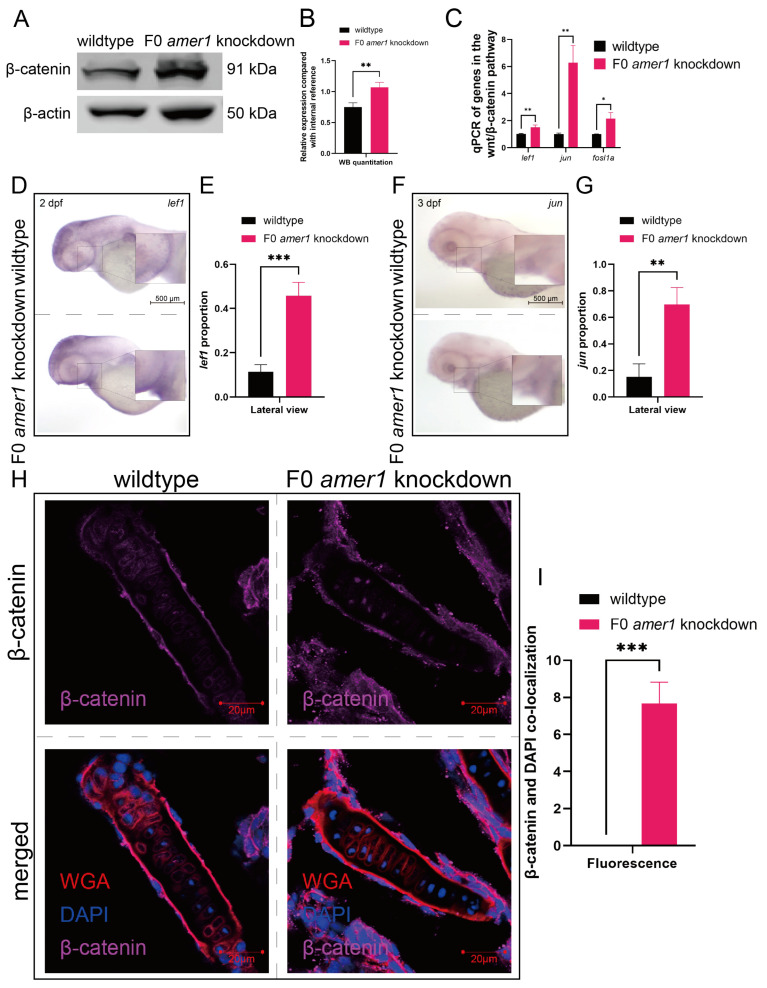
Image
Figure Caption

Figure 8

Expression and localization of components of the wnt pathway in zebrafish embryos. (A,B) Western blotting and quantitation showing that β-catenin levels were higher in amer1 knockdown than in wildtype embryos at 4 dpf. (C) qPCR showing that the levels of expression of lef1, jun, and fosl1a were significantly higher in amer1 knockdown embryos. (DG) Validation of the expression of (D) lef1 and (F) jun by in situ hybridization and their quantitation (E,G) (for quantitative analysis, n = 3 each). (H) Immunofluorescence staining of the ceratohyal cartilage using WGA, β-catenin, and DAPI in wildtype and amer1 knockdown embryo sections at 4 dpf. (I) Manual counting of co-localization of β-catenin with cell nuclei (n = 3 each). * p < 0.05; ** p < 0.01; *** p < 0.001.

Acknowledgments
This image is the copyrighted work of the attributed author or publisher, and ZFIN has permission only to display this image to its users. Additional permissions should be obtained from the applicable author or publisher of the image. Full text @ Int. J. Mol. Sci.