
|
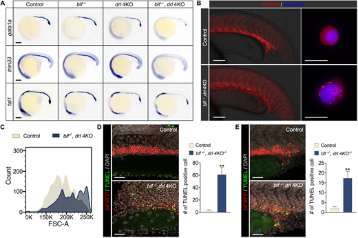
Fig. 3 blf and drl cluster genes are essential for establishing the erythroid cell differentiation program. (A) RNA in situ hybridization of gata1a, trim33 and tal1 expression in 18-somite embryos. Lateral view with head to left. For gata1a WISH, n=37, 35, 33 or 36. For trim33 WISH, n=40, 38, 37 or 32. For tal1 WISH, n=31, 25, 40 or 37. Listed sample sizes relate to each group shown from top to bottom. Scale bars: 100 μm. (B) Fluorescence micrographs of control (blf+/−) and blf−/−; drl 4KO embryos and sorted RFP+ cells at 22 hpf. Scale bars: 200 μm (left); 10 μm (right). (C) Histograms of forward scatter showing approximate cell size distributions of the sorted RFP+ cells. (D,E) Left: Cell death in the intermediate cell mass (D) and in EMPs (E) assessed with TUNEL staining at 22 hpf. Scale bars: 100 μm. Right: Quantification of TUNEL-positive cells counted on a 100 μm×250 μm field for inner cell mass and 125 μm×125 μm field for EMPs. Data are mean±s.e.m. **P<0.01. Data were collected for 8-12 samples per experiment group.