
Image
|
Figure Caption
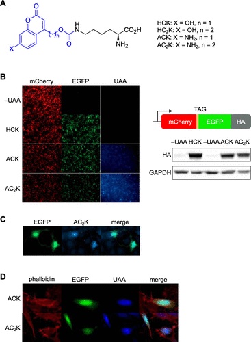
Fig. 1 Incorporation of ACK and AC2K into proteins in mammalian cells. (A) Chemical structures of HCK, HC2K, ACK, and AC2K. The photolabile group/fluorophore is colored blue. (B) Confirmation of ACK and AC2K incorporation into a reporter construct through fluorescence imaging in HEK293T cells (10× magnification) and western blot. (C) Expression of NLS-EGFP-AC2K in live NIH 3T3 cells (20× magnification). (D) Fixed HeLa cells expressing NLS-EGFP-ACK (top row, 40×) and NLS-EGFP-AC2K (bottom row, 63×). Cells are counterstained with rhodamine–phalloidin.
Acknowledgments
This image is the copyrighted work of the attributed author or publisher, and
ZFIN has permission only to display this image to its users.
Additional permissions should be obtained from the applicable author or publisher of the image.
Full text @ ACS Chem. Biol.