
Fig. 5
- ID
- ZDB-IMAGE-230828-99
- Publication
- Domingues et al., 2023 - Cholesteryl hemiazelate Identified in Cardiovascular Disease Patients Causes in vitro and in vivo Inflammation
- All Figures
- Figures for Domingues et al., 2023
|
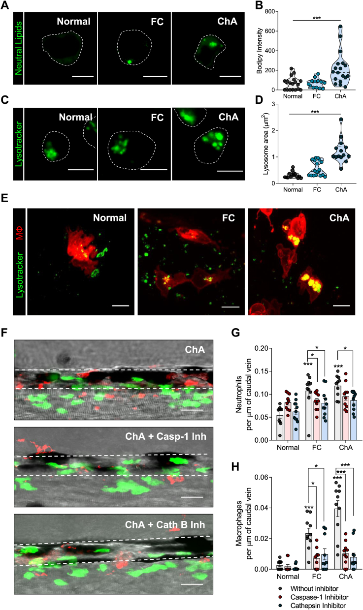
Fig. 5
Inflammasome and cathepsin-B inhibition decrease ChA-induced myeloid cell infiltration. A–D: Zebrafish larvae were fed for 10 days with normal, 4% FC-enriched, or 3% ChA-enriched diets, and macrophages were isolated by fluorescence-activated cell sorting (FACS). A: Representative confocal images of macrophages stained with BODIPY 493/503 to visualize neutral lipids. B: Quantification of neutral lipid accumulation on isolated macrophages. C: Representative confocal images of macrophages stained with LysoTracker to visualize lysosomes/acidic organelles. Scale bars represent 5 μm. D: Quantification of the lysosome area on isolated macrophages. In B and D, the results are the mean ± SEM of two independent experiments (at least 10 cells were analyzed per condition); ∗∗∗