
Fig. 2.
- ID
- ZDB-IMAGE-230503-4
- Genes
- Publication
- Truong et al., 2023 - PRDM1 DNA-binding zinc finger domain is required for normal limb development and is disrupted in split hand/foot malformation
- All Figures
- Figures for Truong et al., 2023
|
Fig. 2.
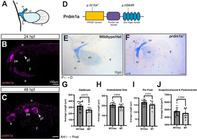
prdm1a−/− zebrafish mutants have hypoplastic pectoral fins. (A) Cartoon of pectoral fin bud at 4 dpf. (B,C) Lateral view of whole-mounted embryos after hybridization chain reaction (HCR) for prdm1a at (B) 24 (n=3) and (C) 48 hpf (n=28). Anterior is to the left. Images are maximum projections of the whole embryo. Arrowheads point to the pectoral fin bud. Scale bars: 100 µm. (D) Schematic of Prdm1a protein. The prdm1am805/m805 allele causes a premature stop codon in the SET domain and is a presumed null mutation (p.W154*). The hypomorphic prdm1atp39/tp39 allele is a missense mutation in the second zinc finger (p.H564R). (E,F) Representative images of Alcian Blue-stained pectoral fins for (E) WT/heterozygous (n=18) and (F) prdm1a−/− mutants (n=9) at 4 dpf. Scale bars: 50 µm. (G-J) The average lengths of the (G) cleithrum, (H) endoskeletal disk and (I) fin fold and (J) the average area of the scapulocoracoid/postcoracoid were measured. Each dot represents one independent biological replicate. Averages were compared with an unpaired, two-tailed independent Student's t-test. prdm1a−/− mutants had a shorter cleithrum (P=0.0173), endoskeletal disk (P=0.0816) and fin fold (P=0.0374). Error bars represent the mean±s.d. The representative images in E and F are also shown in Fig. S1, where the WT and heterozygous fins are analyzed separately. Ant, anterior; cl, cleithrum; D, distal; e, eye; ed, endoskeletal disk; ff, fin fold; Het, heterozygous; hpf, hours post fertilization; MT, prdm1a−/− mutant; n, neurons; P, proximal; pa, pharyngeal arches; pc, postcoracoid; pf, pectoral fin; Post, posterior; sc, scapulocoracoid; WT, wild-type; y, yolk.