
Figure 2
- ID
- ZDB-IMAGE-221226-329
- Genes
- Antibodies
- Publication
- Weeks et al., 2022 - Embryonic alcohol exposure disrupts the ubiquitin-proteasome system
- All Figures
- Figures for Weeks et al., 2022
|
Figure 2
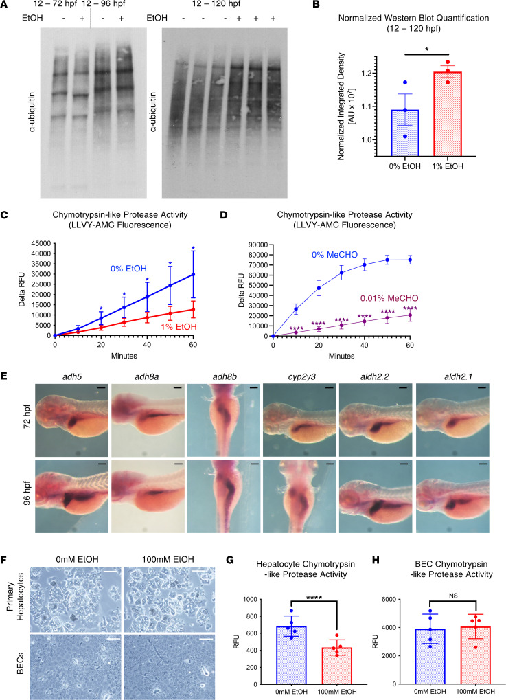
(A) Western blot analysis of ubiquitylated protein and the 20S proteasome after exposure to 0% or 1% EtOH. See Supplemental Figure 2A for loading controls. (B) ImageJ quantification of normalized ubiquitylated protein levels in embryos treated with 0% and 1% EtOH (12–120 hpf; 1-sided unpaired t test, *P ≤ 0.05; n = 3). (C and D) Proteasome activity assay in protein extracts of from whole homogenized 5 dpf larvae. Chymotrypsin-like proteasome activity is impaired by 1% EtOH exposure (12 hpf–5 dpf) and 0.01% MeCHO exposure (104–120 hpf; *P < 0.05, ****P ≤ 0.0001, 2-sided t test per time point; n = 5). (E) Time course ISH for EtOH and MeCHO metabolism genes. For most genes, expression after 72 hpf is noted in the liver and intestine. (F) Confocal imaging of 2D-plated hepatic and biliary epithelial cell (BEC) organoids after 24 hours of treatment with 0 or 100mM EtOH. (G and H) Exposure to 100 mM EtOH impairs chymotrypsin-like proteasome activity in 2D hepatic organoids but not BECs (****P < 0.0001, 2-sided t test; n = 5). Scale bars: 100 μm. For B, G, and H, data represent mean ± SEM. For C and D, data represent mean ± SD.