
|
Figure 2
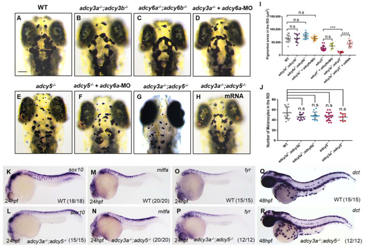
adcy3a-/- and adcy5-/- double mutants show high melanosome aggregation. (A–H) adcy3a-/-;adcy3b-/- (B) and adcy6a-/-;adcy6b-/- (C) double mutants as well as adcy6a-MO-injected adcy3a-/- mutants (D) display normal distribution of melanosomes as that of WT embryos (A) under dark conditions at 4dpf. adcy6a-MO injected adcy5-/- mutants (F) exhibit comparable pigmentation as adcy5-/- (E). adcy3a-/-;adcy5-/- double mutants (G) develop a higher degree of aggregation of melanosomes than adcy5-/-, while co-injection of adcy3a and adcy5 mRNA can significantly rescue the defects in melanosome aggregation in adcy3a-/-;adcy5-/- double mutants (H). Scale bar = 100 μm in (A–H). (I) Pigmentation coverage shows no difference among wild-type embryos, adcy3a-/-;adcy3b-/-, adcy6a-/-;adcy6b-/-, and adcy3a-/- mutants injected with adcy6a-MO, as well as between adcy5-/- and adcy5-/- mutants injected with adcy6a-MO. adcy3a-/-;adcy5-/- double mutants have significantly reduced pigmentation areas compared to adcy5-/-, which can be rescued by co-injection of adcy3a and adcy5 mRNA (Student’s t-test, *** p < 0.001, **** p < 0.0001, n.s., not significant, p > 0.05). (J) Statistical analyses show that the number of melanocytes in adcy5-/- single mutants, and adcy3a-/-;adcy3b-/-, adcy6a-/-;adcy6b-/- and adcy3a-/-;adcy5-/- double mutants is comparable with that of WT embryos (Student’s t-test, n.s., not significant, p > 0.05). (K–R) WISH analyses showed the expression levels of sox10, mitfa, tyr, and dct are normal in adcy5-/-;adcy3a-/- compared to WT embryos.