
|
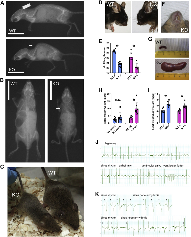
Fig. 7
Phenotypic assessment of Spred2 KO mice
(A) Soft X-ray images showing skeletal abnormalities in a Spred2−/− (KO) mouse compared to an age-matched WT littermate. Arrow labels kyphosis.
(B) X-ray images documenting scoliosis (arrow) in a KO mouse compared to an age-matched WT littermate.
(C) Representative photograph of male littermates at the age of 6 weeks showing growth retardation of a KO mouse (left) compared to an age-matched control animal (right).
(D) Comparison of the head shape of male littermates at the age of 6 months.
(E) Quantification of skull lengths taken from X-ray images (n = 6, each group; ∗p < 0.05).
(F) Representative photograph showing misaligned incisors in KO mice.
(G) Examples of spleens of mice at the age of 12 months showing a dramatic increase in spleen size of a KO mouse.
(H) Quantification of spleen to body weight ratios in young (6–8 weeks) and old (12 months) mice (n = 8 in each group; ∗p < 0.05).
(I) Comparison of heart weight to body weight ratios (n = 6 mice in each group; ∗p < 0.05).
(J) Examples of ventricular arrhythmias in KO mice at the age of 12 months.
(K) Examples of supraventricular arrhythmias in KO mice at the age of 12 months.