
Fig. 3
- ID
- ZDB-IMAGE-221119-3
- Publication
- Shen et al., 2022 - The RNA demethylase ALKBH5 promotes the progression and angiogenesis of lung cancer by regulating the stability of the LncRNA PVT1
- All Figures
- Figures for Shen et al., 2022
|
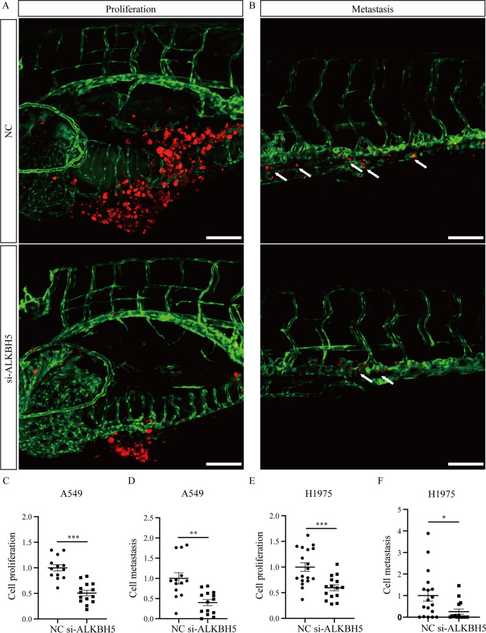
Fig. 3 Knockdown of ALKBH5 inhibits the proliferation and metastasis of lung cancer cells in vivo. A, B The CM-DiI-positive signals in the yolk and trunk of zebrafish were imaged by confocal microscopy at 4 days postinjection of A549 cells transfected with si-ALKBH5 or NC. The red signals indicate the cancer cells labeled CM-DiI. The green signals indicate the blood vessels that expressed EGFP. The white arrows represent the metastatic cells. Scale bar: 100 μm. C, D Statistical analysis of the proliferation and metastasis of A549 cells after knockdown of ALKBH5. NC: n = 13, si-ALKBH5: n = 14. E, F Statistical analysis of the proliferation and metastasis of H1975 cells after knockdown of ALKBH5. NC: n = 13, si-ALKBH5: n = 14. *: P < 0.05, **: P < 0.01, ***: P < 0.001