
FIGURE 1
- ID
- ZDB-IMAGE-220612-1
- Genes
- Publication
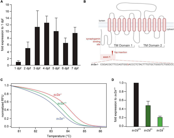
- Zhang et al., 2022 - Connectivity Mapping Using a Novel sv2a Loss-of-Function Zebrafish Epilepsy Model as a Powerful Strategy for Anti-epileptic Drug Discovery
- All Figures
- Figures for Zhang et al., 2022
|
FIGURE 1