
Fig. 8
- ID
- ZDB-IMAGE-220527-29
- Antibodies
- Publication
- Tchivelekete et al., 2022 - Evaluation of okadaic acid toxicity in human retinal cells and zebrafish retinas
- All Figures
- Figures for Tchivelekete et al., 2022
|
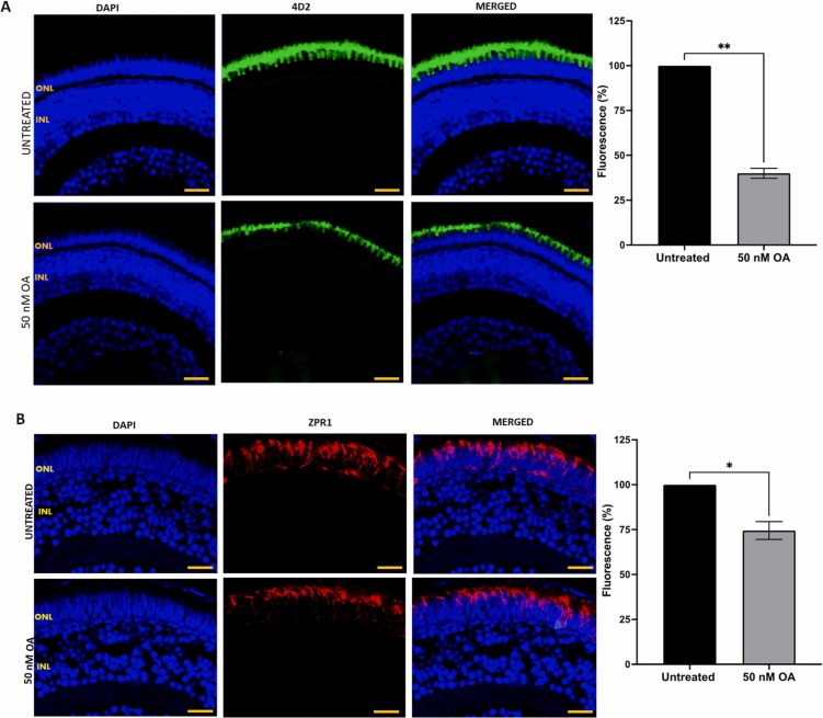
Fig. 8 Fig. 8. (A) Detection of rod cells visualization by immunostaining with anti-rhodopsin 4D2 antibody in zebrafish embryos treated with or without 50 nM OA from 24 to 120 hpf. (B) Cone cells were detected by immunostaining with ZPR-1 antibody in retinal sections of zebrafish embryos treated with or without 50 nM OA from 24 to 120hpf. Embryos exposed to OA illustrated a reduction in fluorescence intensity. Five images were taken from untreated and treated zebrafish retinal sections and quantified using Image J software. Data was presented as mean ± SEM (n = 5). * p < 0.05; * * p < 0.01. Scale bar, 20 µm.