IMAGE

Fig. 3

ID
ZDB-IMAGE-220509-13
Genes
Source
Figures for Amanda et al., 2022
Image
Figure Caption

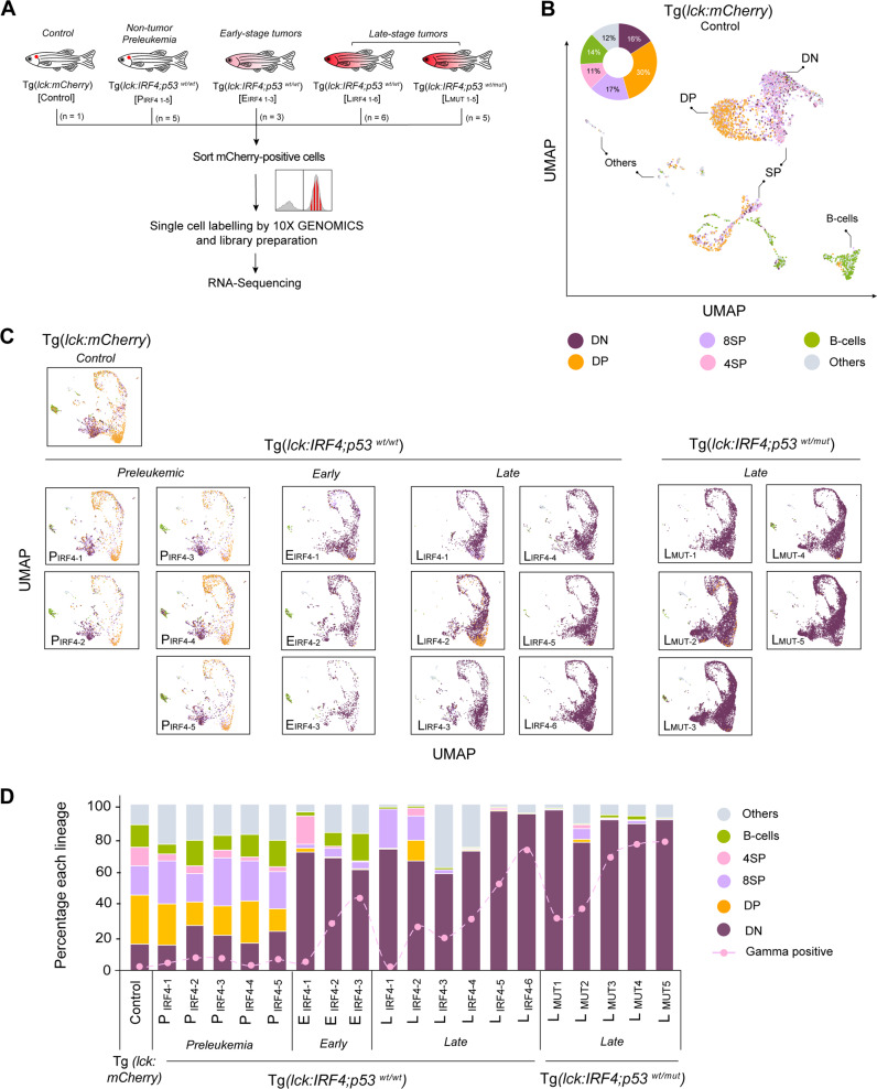
Fig. 3 Identification of multiple cell populations derived from different stages of lymphocyte differentiation.

A Schematic diagram of the sample preparation for scRNA-seq analysis. B UMAP plot showing T and B lymphocytes from one control zebrafish. We first selected T and B-cell clusters and then performed a subclustering analysis. Six populations were then defined based on marker gene expression. C UMAP plots showing aggregated cells from 20 independent zebrafish. Six populations were annotated using the same criteria used in (B). D Bar charts showing the percentage of different cell populations in each sample. Source data are provided as a Source Data file.

Figure Data
Acknowledgments
This image is the copyrighted work of the attributed author or publisher, and ZFIN has permission only to display this image to its users. Additional permissions should be obtained from the applicable author or publisher of the image. Full text @ Nat. Commun.