
|
L
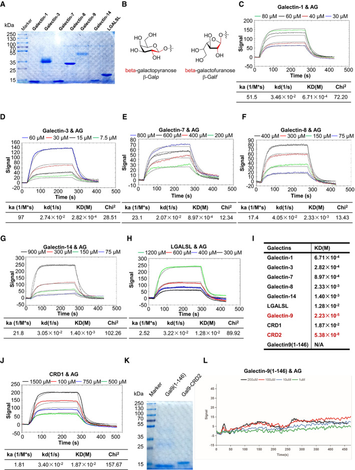
Coomassie blue staining of galectin‐1, galectin‐3, galectin‐7, galectin‐8, galectin‐9, galectin‐14, and galectin‐related protein (LGALSL) post–SDS–PAGE analysis. Chemical structures and conformations of β‐galactofuranoside and β‐galactopyranoside. SPR assay of interactions of AG with indicated galectins including galectin‐1 (C), galectin‐3 (D), galectin‐7 (E), galectin‐8 (F), galectin‐14 (G), LGASL (H), and a summary table of KD (I). Curve fittings to a 1:1 Langmuir‐binding model calculated with TraceDrawer are shown as smooth black lines. The binding affinity of galectin‐9 and CRD2 to AG is highlighted in (I) in red. SPR assay of interactions of AG with CRD1 of galectin‐9. Coomassie blue staining of galectin‐9(1–146) and CRD2 of galectin‐9 post–SDS–PAGE analysis. SPR assay of interactions of AG with galectin‐9(1–146).