
Figure 7
- ID
- ZDB-IMAGE-211201-206
- Genes
- Publication
- Schmitner et al., 2021 - Differential Responses of Neural Retina Progenitor Populations to Chronic Hyperglycemia
- All Figures
- Figures for Schmitner et al., 2021
|
Figure 7
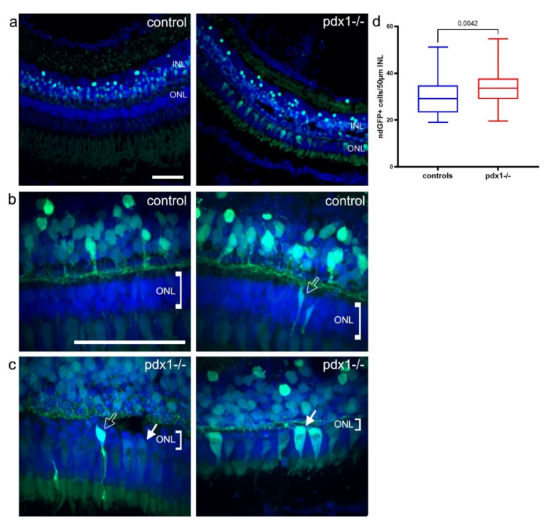
Photoreceptors are restored from neurod-expressing progenitors. (a–c) In neurod:GFP transgenics, strong GFP is detected mainly in the INL in controls, and in the INL and ONL of pdx1 mutants (a). In controls, neurod:GFP labeled rod-shaped cells in the ONL were observed. In pdx1−/− mutants, neurod:GFP labeled rod and cone shaped cells in the ONL in most mutants (b,c, see Table 2). Sections were counterstained with DAPI (blue). A significant increase in GFP positive cells in the INL was detected in pdx1−/− mutants (d). Box plot extends from 75% to 25%, whiskers showing minimum and maximum, line indicates median, n = 10 controls, 9 pdx1−/−. Scale bar: 50 µm. (INL, inner nuclear layer; ONL, outer nuclear layer).