
|
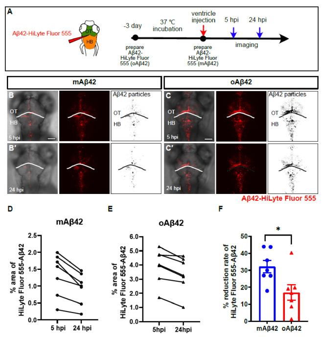
Figure 1
Monomeric Aβ42 peptides are more efficiently cleared from the brain than oligomeric Aβ42. (A) A schematic diagram of experimental setup. Fluorescently labeled Aβ42 prepared at different time points were injected into the brain followed by in vivo imaging at 5 hpi and 24 hpi. (B,C′) Distribution of injected, fluorescently labeled Aβ42 (mAβ42 or oAβ42) in the brains of 3 dpf larvae at 5 hpi (B,C) and 24 hpi (B′,C′) (left, overlays with brightfield; middle, HiLyte Fluor 555; right, thresholded images of Aβ42 fluorescence). (D,E) Quantification of the area fraction (%) occupied by Aβ42 fluorescence within square unit (512 × 512 pixels) at different time points. (F) Clearance rate of mAβ42 and oAβ42 between 5 hpi and 24 hpi by fluorescence quantification. Two-tailed unpaired t-test, p = 0.023. Data are presented as mean ± SEM. N = 7 per group. Data are representative of at least three independent experiments. dpf, days post fertilization; hpi, hours post injection; HB, hindbrain; OT, optic tectum, Scale bars = 100 μm. * p < 0.05.