
Fig 1
- ID
- ZDB-IMAGE-211116-10
- Antibodies
- Publication
- Bühler et al., 2021 - Histone deacetylase 1 controls cardiomyocyte proliferation during embryonic heart development and cardiac regeneration in zebrafish
- All Figures
- Figures for Bühler et al., 2021
|
Fig 1
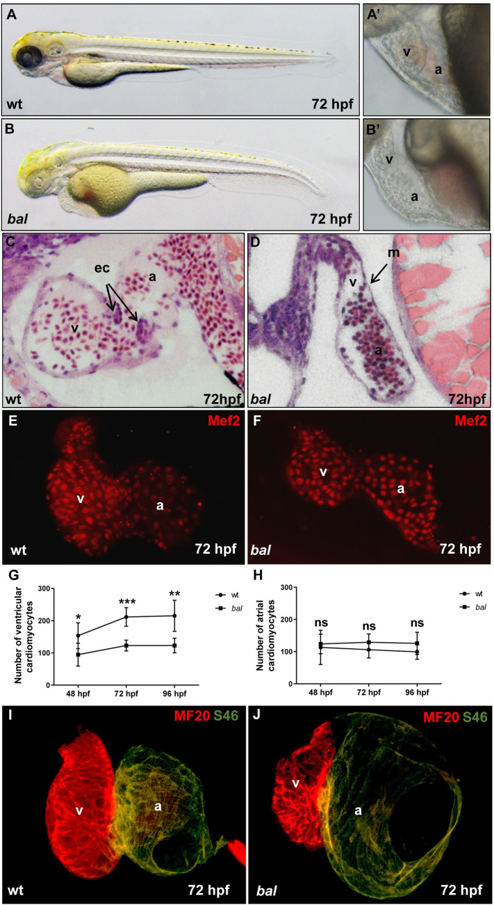
(A-B) Lateral view of 72 hpf wild-type sibling (wt) (A) and bal embryo (B). (A’, B’) Close-up of the heart region of wt (A’) and bal mutants (B’) displayed small ventricle of bal mutants. (C, D) Histological sections showed normal morphology of the wt heart (C) and a monolayered myocardium and small ventricle in the bal mutant heart. M, myocardium; ec, endocardial cushions; v, ventricle; a, atrium. (E, F) Embryonic zebrafish hearts stained with antibodies against MEF-2 to visualize cardiomyocyte nuclei. Cardiomyocyte (CM) numbers appear reduced in bal mutants compared to wild-type siblings. (G) Ventricular cardiomyocyte numbers at 48, 72 and 96 hpf, were significantly decreased in bal (48 hpf wt: 153.37 ± 39.91, n = 7 and bal: 94.71± 35.22, n = 8, p = 0.014; 72 hpf wt: 211.42 ± 28.81, n = 7 and bal: 122.85 ± 16.88, n = 7, p = 0.001; 96 hpf wt: 215.20 ± 48.35, n = 10 and bal 122.83 ± 22.83, n = 6, p = 0.001). Error bars indicate s.d., *p < 0.05, **p < 0.01, ***p < 0.001, ns, not significant. (H) Atrial CM numbers were unaltered (48 hpf p = 0.332, 72 hpf p = 0.135, 96 hpf p = 0.147). (I, J) Immunofluorescent (IF) staining against meromyosin (MF20) (red) and atrial-specific myosin (S46) (green) demonstrated normal chamber specification of bal hearts.