IMAGE

Fig. 2

ID
ZDB-IMAGE-211009-56
Source
Figures for Fragale et al., 2021
Image
Figure Caption

Fig. 2

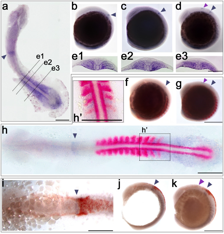
atoh8, myoD and crestin expression in early segmentation stages of development (13–14 hpf). aeatoh8 ISH; fhatoh8/myoD double ISH. ikatoh8/crestin double ISH. a Flat mount of 13.5-hpf embryo. b 13-hpf embryo showing first appearance of atoh8 in the developing hindbrain (dark arrowhead). c 13.5-hpf embryo. d Embryo at 14 hpf; pink arrowhead indicates the appearance of the second domain at the midbrain/hindbrain boundary. e1e3 Sections through the 13-hpf embryo at the levels indicated in a. f, g Embryos at 13 hpf and 14 hpf, respectively, double-stained for atoh8 and myoD. h Flat mount of embryo at 14 hpf double-stained for atoh8 and myoD. i, j Flat mount and embryo showing double labelling for crestin and atoh8 at 13 hpf. k Embryo at 14 hpf double-stained for crestin and atoh8. a, h’ Dorsal view, anterior to the top. bd, f, g, j, k lateral view, anterior to the top, dorsal to the right. h, i dorsal view, anterior to the left. Scale bars: whole embryos 200 µm, flat mounts a, h, i 200 µm; h’ 50 µm; transverse sections: 100 µm

Acknowledgments
This image is the copyrighted work of the attributed author or publisher, and ZFIN has permission only to display this image to its users. Additional permissions should be obtained from the applicable author or publisher of the image. Full text @ Histochem. Cell Biol.