
Fig. 2
- ID
- ZDB-IMAGE-210927-6
- Publication
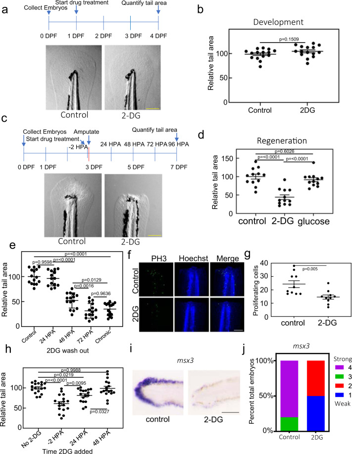
- Sinclair et al., 2021 - The Warburg effect is necessary to promote glycosylation in the blastema during zebrafish tail regeneration
- All Figures
- Figures for Sinclair et al., 2021
|
Fig. 2