
IMAGE
Figure 3
- ID
- ZDB-IMAGE-210821-15
- Publication
- Del Bene et al., 2007 - In vivo validation of a computationally predicted conserved Ath5 target gene set
- All Figures
- Figures for Del Bene et al., 2007
Image
|
Figure Caption
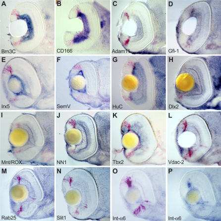
Figure 3
(A–P) Embryos are sectioned transversally at the level of the optic nerve. Dorsal is to the top. Abbreviations of target genes are indicated in
Acknowledgments
This image is the copyrighted work of the attributed author or publisher, and
ZFIN has permission only to display this image to its users.
Additional permissions should be obtained from the applicable author or publisher of the image.
Full text @ PLoS Genet.