
Figure 4
- ID
- ZDB-IMAGE-210719-4
- Publication
- Adams et al., 2021 - A two-site flexible clamp mechanism for RET-GDNF-GFRα1 assembly reveals both conformational adaptation and strict geometric spacing
- All Figures
- Figures for Adams et al., 2021
|
Figure 4
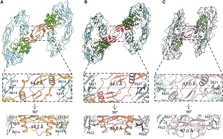
Different GFL ligands establish a conserved spacing between RET CRD-CRD pairs in their respective ternary complexes
(A) Separation between the Cα of E613 (equivalent to E620 of hRET) from both molecules of zRETECD within the zRGα1a structure.
(B) Equivalent distance between the Cα E620 from both molecules of hRETECD from the hRETECD-NRTN-GFRα2 (PDB:
(C) Equivalent separation between the Cα E620 from the two molecules of hRETECD from the hRETECD-GDF15-GFRAL (PDB:
Reprinted from Structure (London, England : 1993), 29(7), Adams, S.E., Purkiss, A.G., Knowles, P.P., Nans, A., Briggs, D.C., Borg, A., Earl, C.P., Goodman, K.M., Nawrotek, A., Borg, A.J., McIntosh, P.B., Houghton, F.M., Kjær, S., McDonald, N.Q., A two-site flexible clamp mechanism for RET-GDNF-GFRα1 assembly reveals both conformational adaptation and strict geometric spacing, 694-708.e7, Copyright (2021) with permission from Elsevier. Full text @ Structure