
Figure 2
- ID
- ZDB-IMAGE-210719-2
- Publication
- Adams et al., 2021 - A two-site flexible clamp mechanism for RET-GDNF-GFRα1 assembly reveals both conformational adaptation and strict geometric spacing
- All Figures
- Figures for Adams et al., 2021
|
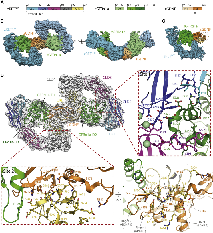
Figure 2
Cryo-EM structure of the zRETECD-zGFRα1aD1-3-zGDNFmat. (zRGα1a) complex
(A) Schematic of zRETECD, zGFRα1aD1-3, and zGDNFmat., color coded according to
(B) Orthogonal views of the reconstituted zRGα1a complex cryo-EM map, projecting down the approximate molecular dyad or perpendicular to it. The cryo-EM map is segmented and colored by protein, with zRETECD cyan, zGFRα1aD1-3 green, and zGDNFmat. orange.
(C) Symmetry-expanded map of zRGα1a half-complex, with the map segmented and colored by protein as in (B).
(D) The final model of the zRGα1a complex built into the C2 symmetry map, colored light gray. The domains are colored as in
Reprinted from Structure (London, England : 1993), 29(7), Adams, S.E., Purkiss, A.G., Knowles, P.P., Nans, A., Briggs, D.C., Borg, A., Earl, C.P., Goodman, K.M., Nawrotek, A., Borg, A.J., McIntosh, P.B., Houghton, F.M., Kjær, S., McDonald, N.Q., A two-site flexible clamp mechanism for RET-GDNF-GFRα1 assembly reveals both conformational adaptation and strict geometric spacing, 694-708.e7, Copyright (2021) with permission from Elsevier. Full text @ Structure